Vitreoretinal Surgery

Vitreoretinal Surgery
Vitrectomy
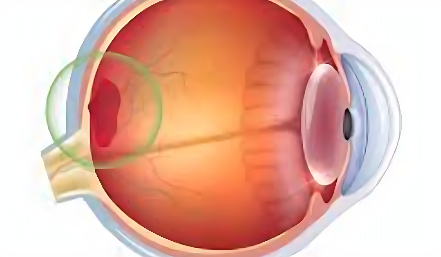
Vitrectomy is a type of eye surgery that involves the removal of the vitreous, a gel-like substance that fills the inside of the eye. It is also known as pars plana vitrectomy.
It is used to treat a variety of eye conditions, including:
• Diabetic retinopathy: This is a complication of diabetes that can damage the blood vessels in the retina.
• Age-related macular degeneration (AMD): This is a leading cause of vision loss in adults over 50.
• Retinal detachment: This is a condition in which the retina separates from the back of the eye.
• Macular hole: This is a small hole in the center of the retina that can cause vision loss.
• Epiretinal membrane: This is a thin layer of scar tissue that forms on the surface of the retina and can cause blurred vision.
• Vitreous hemorrhage: This is a condition in which the vitreous bleeds.
Vitrectomy is performed to treat these conditions by removing the vitreous and replacing it with a clear solution. This allows the surgeon to access and repair the underlying eye condition.
Most people make a full recovery from vitrectomy within a few weeks. However, it is important to follow the surgeon’s instructions carefully after surgery to prevent complications.
If you have a vitrectomy, you will need to avoid strenuous activities for a few weeks after surgery. You will also need to use eye drops to prevent infection and inflammation.
Intravitreal Injections
An intravitreal injection is a surgery to inject a medication into the vitreous, the clear gel that fills the inside of the eye. Intravitreal injections are used to treat a variety of eye conditions, including:
• Age-related macular degeneration (AMD): AMD is a leading cause of vision loss in adults over 50. It is caused by damage to the macula, the central part of the retina that is responsible for sharp central vision.
• Diabetic retinopathy: Diabetic retinopathy is a complication of diabetes that can damage the blood vessels in the retina. It is a leading cause of blindness in adults.
• Retinal vein occlusion (RVO): RVO is a condition that occurs when a blood vessel in the retina is blocked. It can cause vision loss, swelling, and bleeding in the eye.
• Uveitis: Uveitis is an inflammation of the uvea, the middle layer of the eye. It can cause pain, redness, and vision loss.
The procedure takes about 15 minutes. During the injection, the doctor will numb the eye with eye drops. They will then use a sterile needle to inject the medication into the vitreous. The medication will slowly release into the retina over time. Most people need a series of intravitreal injections to treat their condition.